Contents:
- Medical Video: Dental Xrays SAFE? (How often and why for dental health and oral health?) - EP 06 The Dental Drive
- When is X-rays needed?
- Does X-rays have risks?
- Types of chest x-ray examination
- Postero-anterior Projection
- AP (Antero-Posterior) Projection
- Lateral projection
- Preparation must be done before undergoing X-rays
- Conventional radiography without preparation
- Conventional radiography with preparation
Medical Video: Dental Xrays SAFE? (How often and why for dental health and oral health?) - EP 06 The Dental Drive
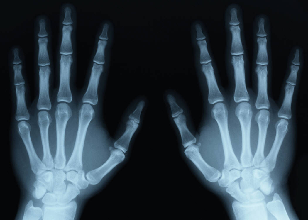
Roentgen or X-ray rays, which in Indonesia are better known as X-rays, were discovered by German physicist Wilhelm Roentgen, precisely on November 8, 1890. These rays are able to penetrate parts of the human body without surgery (non-invasive procedure) so the medical world was greatly helped by this finding. Thanks to his achievements, Roentgen was awarded the Nobel Prize in 1901.
When is X-rays needed?
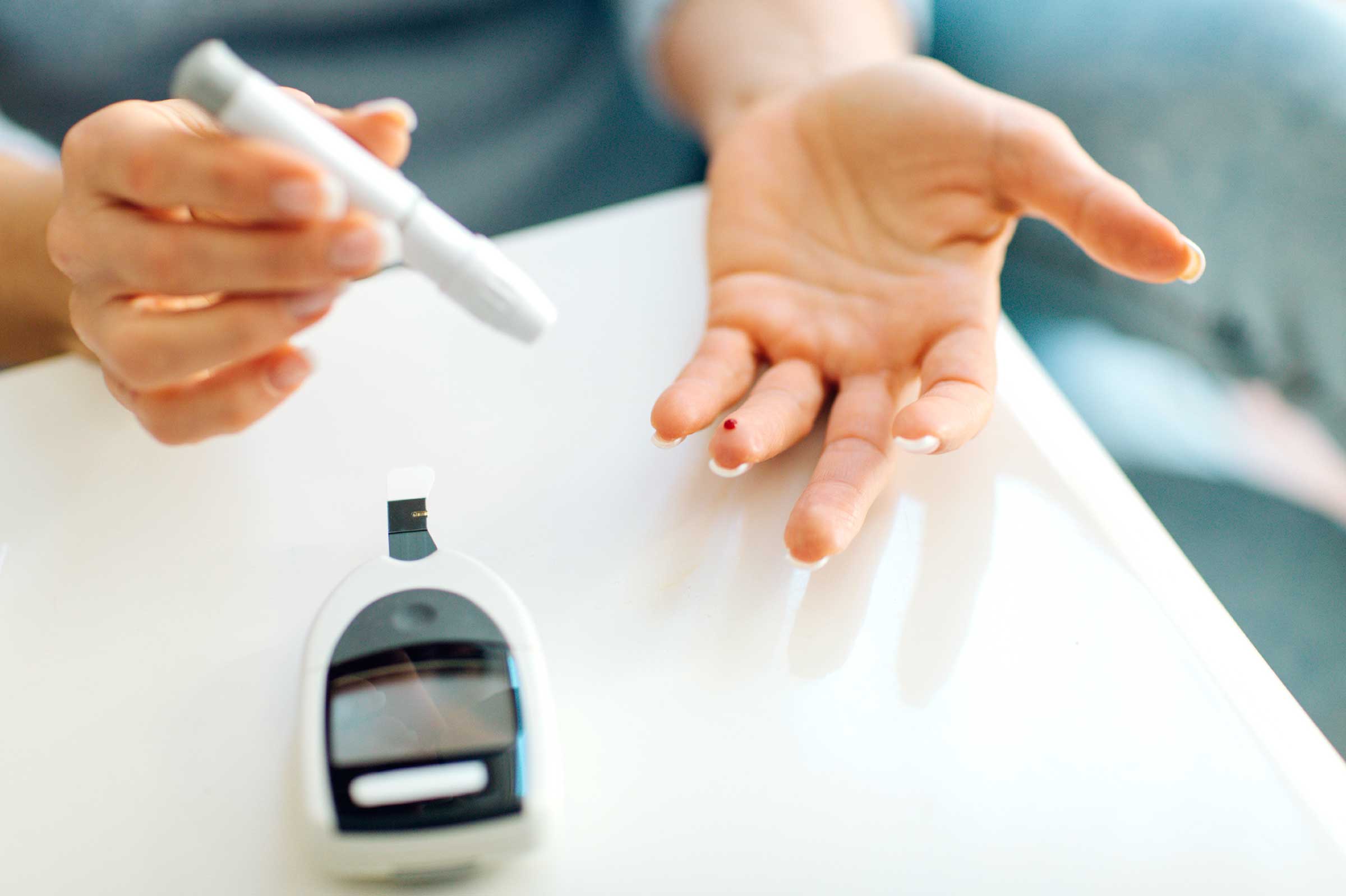
X-ray examination is one of the investigations of establishing a diagnosis in addition to laboratory tests. X-rays are performed to see fractures or fractures, monitor their progress, and determine the type of treatment to be given.
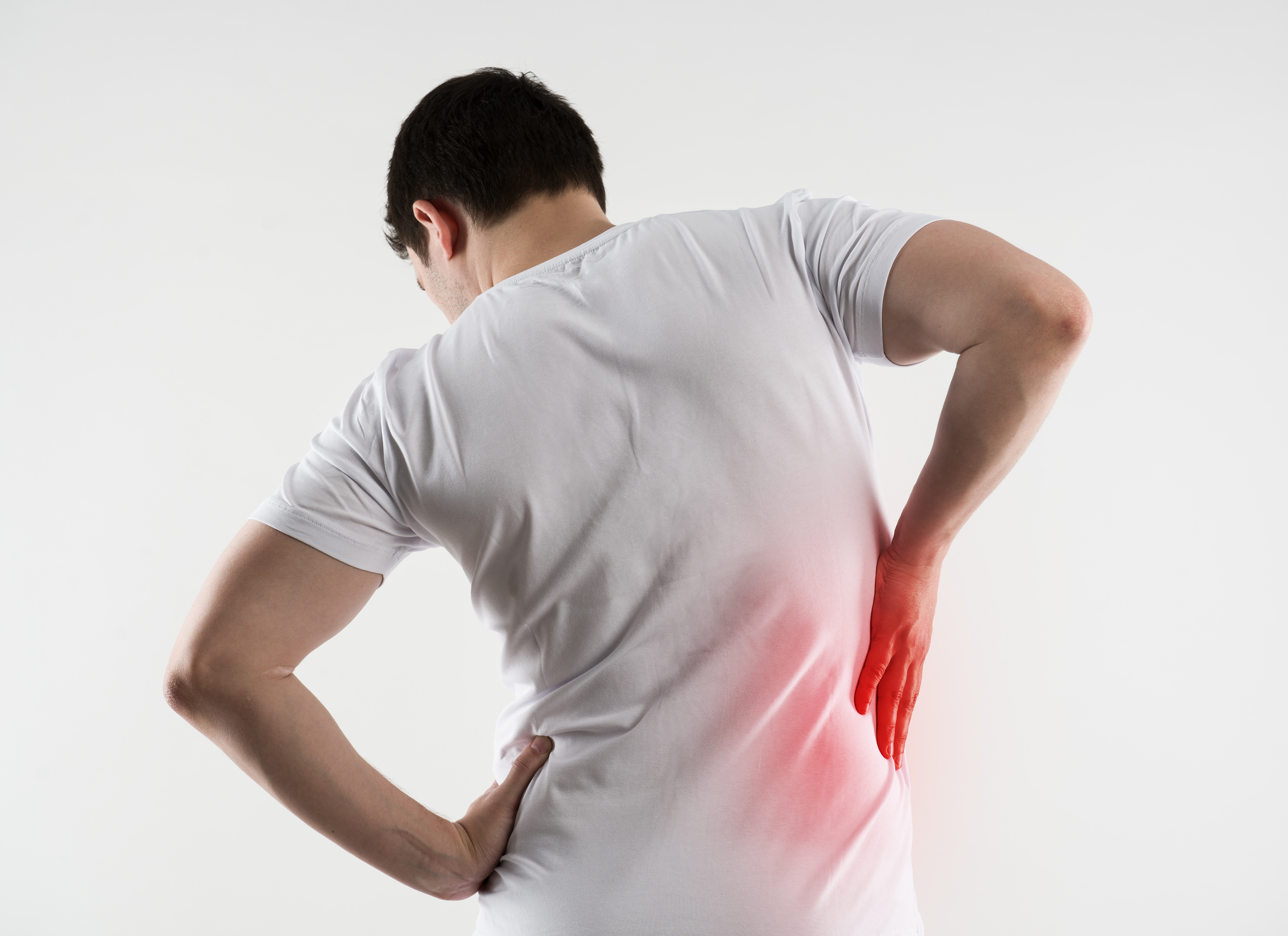
Disease conditions that require x-ray examination for example are arthritis, bone cancer, lung disease, digestive problems, enlarged heart, kidney stones, urinary tract stones, and the occurrence of ingestion of foreign substances.
Does X-rays have risks?
X-rays use only a little radiation, so the amount of light exposure is still considered safe for adults. Unlike the case with the fetus in the womb, so for pregnant women usually carried out radiological examinations with other types that are safer such as MRI.
In addition, there are some X-ray examination conditions that require the ingestion or injection of contrast substances so that the photos of the area you want to see can be clearly illustrated. The commonly used contrast is the type of iodine that some people can cause allergies. Allergic reactions that can occur are redness of the skin, itching, and nausea. In very rare cases anaphylactic shock can occur, severe hypotension, and a heart attack.
Types of chest x-ray examination
Postero-anterior Projection
How to examine chest photographs with a PA (Postero-anterior) projection, namely:
- The light is emitted towards the film through the patient's (posterior) back. Usually, patients will be asked to stand upright with the anterior region (abdomen) attached to the film.
- Hands flank to lift the shoulder blades so that the lung region is not covered.
- The patient is asked to take a deep breath when the beam is fired so that the thoracic cavity can expand optimally, the diaphragm will be pushed into the abdominal cavity (stomach) so that it can produce a picture of the lung / heart like the original. This examination can only be done in the radiology room
AP (Antero-Posterior) Projection
How to examine chest radiographs with AP (Antero-Posterior) projections, namely:
- AP projections can be performed on patients who are supine, seated, or supine, but the angle of the stem is 45 or 90 degrees from the flat plane.
- This procedure is usually performed on patients who cannot move (mobilization) due to various causes, often occurring in postoperative patients.
- The tool used is a photo tool portable.
- AP projection images usually produce poor photo quality compared to PA projections
Lateral projection
How to examine chest radiographs with lateral projections, namely:
- This position is carried out according to the indications of both the right and lateral lateral left
- Usually done if needed to make a diagnosis that is not obtained with other projected photos.
Preparation must be done before undergoing X-rays
Based on the type of preparation, the X-Ray examination is divided into:
Conventional radiography without preparation
Patients can be directly photographed when they arrive.
Conventional radiography with preparation
- Examination of the abdominal organs (stomach) requires fasting for several hours or only eating certain foods so that the intestines can be clearly drawn without any closure from the stool.
- On the urinary tract examination, you will be asked to lie on your back with your hands away from your body. As well as before the examination you will be asked to drink plenty of water or hold urine so that a good picture of the bladder (bladder) can be seen.
- Examination of the chest posterior anterior (PA) projection is done in a standing position, the clothes must be lowered to the waist. You will be asked to hold your breath when the photo is taken.
- If x-rays are done on the skull area, hair clips or ornaments, glasses, and dentures must be removed.
Other technical preparations are as follows:
- Wear comfortable and loose clothing to make it easy to open, but some hospitals will be given dresses to wear.
- Take off jewelry, clocks or metal-containing instruments on the body. If you have metallic implants in the body from a previous operation, immediately report it to the doctor because the implants will block X-ray rays to penetrate into the body.