Contents:
- Medical Video: Journey of Sound to the Brain
- Understanding the anatomy of the human ear
- Outer ear (outer ear)
- Middle ear (middle ear)
- Inner ear (inner ear)
- How can you hear?
Medical Video: Journey of Sound to the Brain
Hearing is one of the abilities in the human ear that supports communication with one another. In addition, the ear also functions to maintain the balance of the body. If your ears experience interference, of course the activities that you do also experience obstacles. To find out more, see the review of the following ear anatomy.
Understanding the anatomy of the human ear
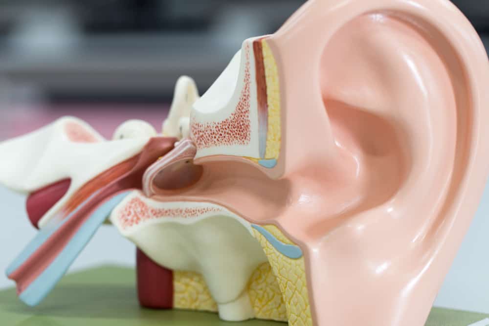
The human ear consists of three parts, namely the outer ear (outer ear), middle ear (middle ear), and finally the inner ear (inner ear). Note the illustration of the ear anatomy based on the following three parts.
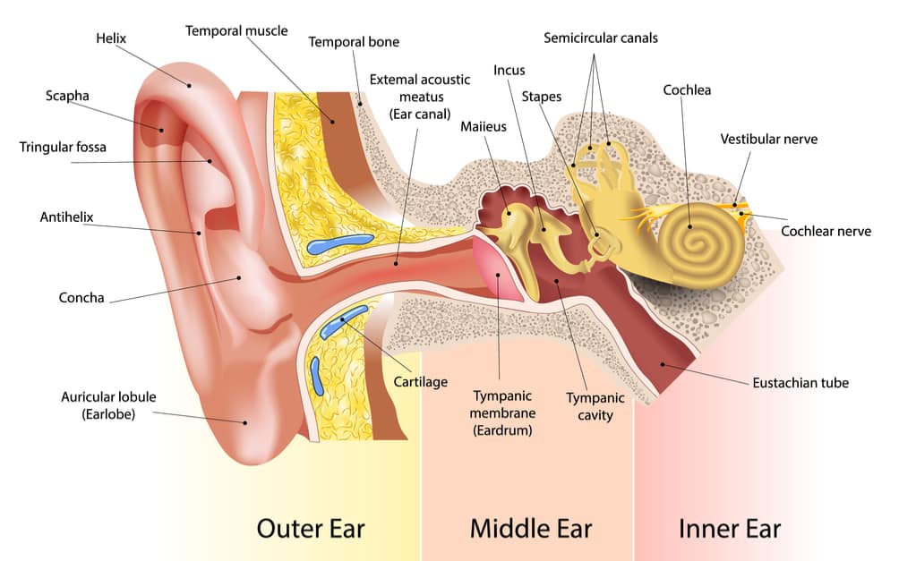
Outer ear (outer ear)
This part of the ear is formed from auricula (auricle) and external auditory canal (ear canal or ear canal) Auricula is formed by elastic cartilage that is firmly attached to sloping skin. It functions to capture sound and localize sound. The auricula forms a basin called concha and the edges are called helices.
The earlobe consists of:
- Helix
- Spiral
- Antiheliks
- Scaphoid fossa
- Fosa triangle
- Antihelical crura
- Antitragus
- Lobule
- Tragus
Ear canal (ear canal) formed by cartilage and temporal bone. The size is about 4 cm from the tragus to the tympanic membrane (tympanic membrane) which is also called the eardrum and curves to form the letter S. The curvature is useful for preventing foreign objects from reaching the tympanic membrane. There is condyle of the mandible at the front of the ear canal bone and mastoid air cells at the end.
There are several sensory nerves in the outer ear, such as the auricular nerve, the occipital nerve, the ariculotemporal nerve, and the auricular branch of the fagus nerve (arnold nerve).
Middle ear (middle ear)
The function of this part of the ear is to deliver sounds that have been collected by the auricula to the inner ear. This part of the ear extends from the cavity to the tympanic membrane to the oval window which consists of the bones of the malleus, incus, and stapes and many complicated walls. For example, the lateral wall, medial wall, tagmental wall, and jugular wall.
The tympanic membrane is thin and semi-transparent which separates the outer ear from the middle ear which consists of the flaccida pars and the pars tensa. The malleous manubrium is firmly attached to the tympanic membrane in the form of a basin called umbo. The higher part of the umbo is what is called the pars flaccida and the rest is called pars tensa.
There are three sensory nerves in the tympanic nerve, namely the auriculotemporal nerve, the arnold nerve, and the tympanic nerve branch. On the surface of the tympanic membrane there are moving bone chains called ossicles, namely malleus (hammer), incus (runway), stapes (stirrups). These bone elements function to deliver and strengthen sound waves up to 10 times stronger than air into the inner ear perilymph.
In addition, there is an eustachian tube that connects the middle ear to the upper part of the esophagus and nose (nasopharynx). Its function is to equalize the air pressure with the movement open and close. Important muscles in the middle ear include the stapedius muscle and the tensor tympani tendon.
The horizontal part of the facial nerve crosses the tympanic cavity. Therefore, if there is paralysis of the nerves or facial muscles, the sound sharpness will be blocked and damage to the inner ear.
Inner ear (inner ear)
This part of the ear is called the labyrinth cavity which functions to help balance and channel sound to the central nervous system. This cavity is formed from the osseous labyrinth, namely a series of temporal bones and a membrane labyrinth (membrane sacs and ducts). The membrane labyrinth also has a cochlear, vestibular, and semicircular (semicircular) component.
Cochlear (cohclea) is an important organ in the inner ear shaped like a snail shell. The shape is like a tube that bends backward as far as 2.5 circles with a cone shape at the end. This section has three chambers, namely the vertibuli scale, the cochlear channel, and the tympanic scale. In this cochlea, there is a cortical organ which functions to convert sound waves into nerve impulses.
Vestibuli are the connecting part between the cochlea and the semicircular channel. It consists of the sac and utricula, the hair cells that maintain the balance of the head's position against the force of gravity when the body is stationary.
While semicircular is a semicircular canal of three different channels, namely the horizontal semicircular canal, the upper vertical semicircular canal, and the rear vertical semicircular canal containing the ampulla. This serves to determine the position of the head when there is a rotational or rotating motion.
How can you hear?
From the ear anatomy, you have learned the parts that make up the ear, namely the outer ear, middle ear, and outer ear. The three parts of the ear become channels of sound from outside to enter and be translated in the brain.
Reporting from Stanford Childrens, the sound from around you is captured by the outer ear in the form of vibrations or waves. Then, the sound is lowered into the ear canal so as to put pressure or blow on the eardrum (tympanic membrane). When the eardrum vibrates, vibrations will be transmitted to the bone ossicles so that vibrations are strengthened and sent to the inner ear.
Once the vibration reaches the inner ear, the vibration will be converted into electrical impulses and sent to the auditory nerve in the brain. The brain then translates this impulse as sound.
After knowing the anatomy of the ear, you must understand that the ear is not only a hearing device, but also maintains balance. This allows you to walk, jump, run without falling. If you feel a disturbance in your ear, immediately check your health to the doctor to get the right diagnosis and treatment.