Contents:
- Medical Video: Clearing Your Chest with Breathing Exercises
- Pulmonary anatomy, complete from the largest to the smallest channel
- 1. Pleura
- 2. Bronchus (Bronci)
- 3. Bronchiole (Bronchioles)
- 4. Alveoli
- Various diseases caused by disruption of lung function
Medical Video: Clearing Your Chest with Breathing Exercises
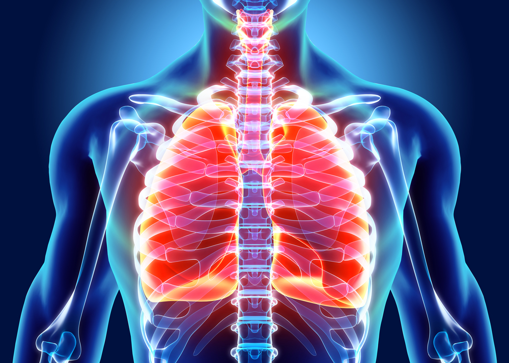
The lungs are organs that are responsible for processing incoming air, separating oxygen from carbon dioxide. This organ consists of two pairs, each of which has different characteristics. Intrigued by the function and what are the parts of the lungs? Come on, get to know more about the anatomy of this human lung.
Pulmonary anatomy, complete from the largest to the smallest channel
Basically, the right and left lungs have different characteristics. For example, from the weight, the left lung of an adult weighs about 325-550 grams and the right lung has a weight of about 375-600 grams.
The left lung consists of two parts (lobes) while the right has three different parts (lobes). Therefore, the right lung has a size and weight greater than that.
To find out more about lung anatomy, see the following reviews and images.
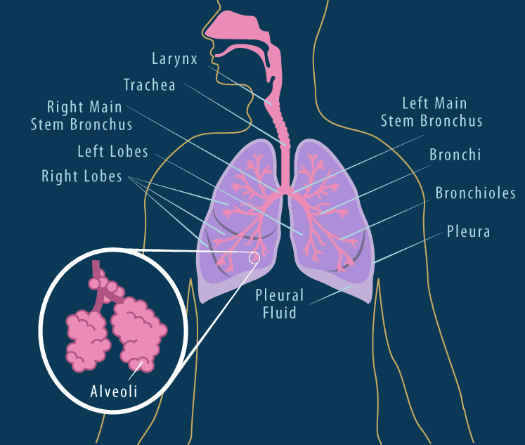
1. Pleura
The pleura is a double-layered thin membrane that lines the lungs. This layer secretes fluid (pleural fluid) called serous fluid which functions to lubricate the inside of the lung cavity so it does not irritate the lung when it expands and contracts while breathing.
2. Bronchus (Bronci)
The bronchus is a branch of the windpipe located after the throat (trachea) before the lungs. The bronchus is an airway that ensures that air enters well from the trachea to the alveoli.
Aside from being an entry and exit air, the bronchi also functions to prevent infection. This is because the bronchi are covered by various types of cells, including cells that are ciliated (hairy) and slimy. These cells will then trap disease-carrying bacteria not into the lungs.
3. Bronchiole (Bronchioles)
Bronchiole is a branch of the bronchi that serves to channel air from the bronchi to the alveoli. In addition, bronchioles also function to control the amount of air entering and leaving during the breathing process.
4. Alveoli
This part of the lung anatomy is the smallest group called the alveolar sac at the end of the bronchiole. Each alveoli is a concave shaped cavity surrounded by many small capillaries.
Its function is as a place for exchanging oxygen and carbon dioxide. The alveoli then absorbs oxygen from the air carried by the bronchioles and drains it into the blood.
After that, carbon dioxide which is a waste product from body cells flows from the blood to the alveoli to be blown out. This gas exchange occurs through the very thin walls of the alveoli and capillaries.
Various diseases caused by disruption of lung function
Here are some diseases that commonly affect the respiratory system due to impaired lung function, namely:
- Bronchitis, namely respiratory diseases that occur due to upper respiratory tract infections and are usually caused by viruses.
- Pneumonia, which is a respiratory disorder that causes the smallest part of the lungs, namely the bronchioles and alveolar tissue to become inflamed.
- Asthma, usually caused by inflammation of the respiratory tract. As a result, the airway swells and narrows, blocking the passage of air that should flow into the lungs.
- Tuberculosis, is a bacterial infection caused by Mycobacterium tuberculosis which attacks and damages the tissues of the human body. These bacteria can be transmitted through the airways. TB usually attacks the lungs, but can also spread to the bones, lymph nodes, central nervous system, heart, and other organs.