Contents:
- Medical Video: Atoms for Health - The Role of Nuclear Techniques in Medicine
- List of medical procedures in Indonesia with nuclear power
- 1. Radionuclear therapy
- 2. Renogram
- 3. PET scan
- 4. Branchytherapy
Medical Video: Atoms for Health - The Role of Nuclear Techniques in Medicine
Hearing the word "nuclear" and "radioactive compounds" certainly makes you shudder in horror. Because maybe you think how terrible the danger of nuclear power is in war. Eits, don't get me wrong. In recent years, nuclear energy has been developed as a supporting material for medical examinations in Indonesia. Indeed, what types of nuclear power-based health checks exist in Indonesia? Come on, see the full review below.
List of medical procedures in Indonesia with nuclear power
1. Radionuclear therapy
During this time, many cancer treatments have been directed at chemotherapy or radiotherapy. In fact, there are other treatment alternatives that are considered effective in treating cancer, namely radionuclear therapy.
Simply put, radionuclear therapy is a medical procedure that utilizes heat from nuclear radiation as a therapy for disease. Radionuclear therapy is useful for treating a number of cancers, including thyroid cancer, nasopharyngeal cancer, lymph node cancer, and neuroblastoma (nerve cell cancer in children).
Just like chemotherapy, this therapy is systemic or reaches the entire body through the bloodstream. But the difference is, radioactive substances in this therapy specifically target cancer cells specifically by damaging the DNA of cancer cells. As a result, cancer cells become more easily controlled and the side effects caused are even less than the effects of chemotherapy.
However, this radionuclear is only available to several hospitals in big cities. The cost that must be spent is fairly large for several therapy sessions.
2. Renogram
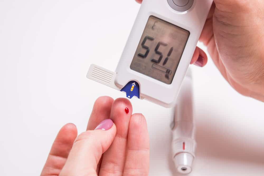
Renogram is a nuclear-based medical examination that is used to map kidney function. This procedure is used to measure and monitor the extent to which a patient's kidneys are working properly.
Before undergoing a renogram, the patient will be asked to empty the bladder first. Patients are allowed to keep wearing their clothes, but are obliged to remove all metal objects that are attached to the body, such as braces, jewelry or belts.
Furthermore, the patient will be asked by the doctor to lie in bed or sit in a special chair. In the patient's chair there is a gamma camera that is parallel to the lower back or the location where the kidney is located.
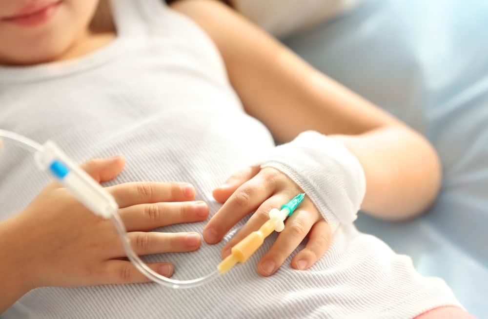
The patient will be injected with a radionuclide in the form of a compound Iodine-131 into a vein in the arm. This radionuclide will flow throughout the patient's body and filtered by kidney organs. The patient only needs to sit for 30 to 60 minutes as long as the gamma camera takes a series of images or images on the patient's kidney.
The advantage of this medical examination is that the patient will not feel any effect. The reason is, the renogram procedure will not emit radiation, but only detects radiation originating from injected radionuclides.
The product produced by the renogram is a graph that shows how quickly radionuclides pass through the kidneys and enter the patient's bladder. If the chart pattern tends to be standard, then the patient's kidney function can be said to be in good condition. Conversely, if there is a chart that deviates from the standard, it can be said that the patient's kidney function has certain problems.
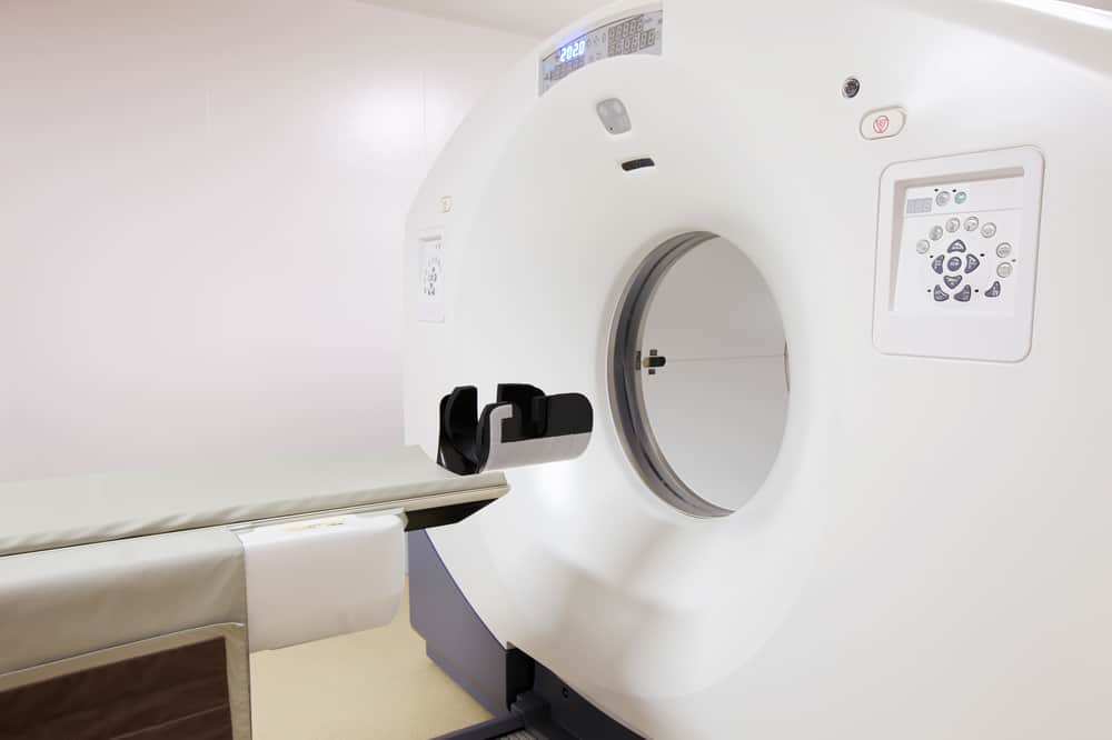
3. PET scan
Another form of utilization of nuclear power in the health sector is scanning Positron Emission Tomography (PET). PET scans are imaging tests with radiation to see cell activity in the body.
This procedure is most often used to investigate epilepsy, Alzheimer's disease, cancer, and heart disease. When PET scans are used to detect cancer, doctors will see how the cancer metabolizes the body and whether the cancer has spread (metastasis) to other organs.
Before undergoing PET scans, patients should not eat any food for 4 to 6 hours before scanning. However, patients still need to consume lots of water to prevent dehydration.
The patient will then be injected with a number of radiotracers, a tracker containing radioactive and natural chemicals such as glucose. This radiotracer will move towards the target cell by using glucose as energy. Because the body needs time to absorb the radiotracer, the patient must wait about an hour before the scan begins. Only then is the patient asked to lie on the surface connected to the PET machine and start scanning.
4. Branchytherapy
Branchytherapy is one medical procedure that utilizes nuclear power. Medical examinations often referred to as local radiation are used to treat a number of cancers, such as brain cancer, breast cancer, cervical cancer, eye cancer, lung cancer, and other types of cancer.
Branchytherapy allows doctors to give higher radiation doses to specific areas of the body. However, the side effects and duration of healing are actually faster than other external radiation.
This medical examination can be done separately or in conjunction with other cancer treatments. For example, branchytherapy is sometimes used to help destroy the remaining cancer cells postoperatively, or it can also be done in conjunction with external beam radiation.
Branchytherapy examination is done by inserting radioactive material directly in the body near the location of the cancer. However, this is influenced by many factors, including the location and severity of the cancer, the overall health condition of the patient, and the purpose of the treatment itself.
This radioactive can be placed on two parts of the body, namely:
1. In the body cavity
During branchytherapy intracavity, a device containing radioactive material will be placed in the body cavity, such as the throat or vagina. This tool can be a tube or cylinder that matches the size of the target body cavity. This set of devices is then positioned by the hands of the radiation therapy team or with the help of the machine to be right at the location of the cancer.
2. In body tissues
During interstitial branchytherapy, a device containing radioactive material will be placed in the body's tissues, such as in the breast or prostate. This tool consists of a needle and a small balloon about the size of rice at the end. CT scans, ultrasound (ultrasound), or other imaging are then used to help direct the device to cancerous tissue and begin scanning.