Contents:
- Medical Video: Anatomy of the Pelvis - for Artists
- Female pelvic anatomy
- Women's pelvic bones
- Pelvic canal
- Size and shape of the pelvis
Medical Video: Anatomy of the Pelvis - for Artists
Every woman has a pelvis that is unique and different from one another. The difference in pelvic organs is influenced by genetics, lifestyle, such as physical activity, eating habits, health conditions, sexual activity, and much more. To find out more about the woman's pelvis, let's look at the following review about the anatomy of the female pelvis and its function.
Female pelvic anatomy
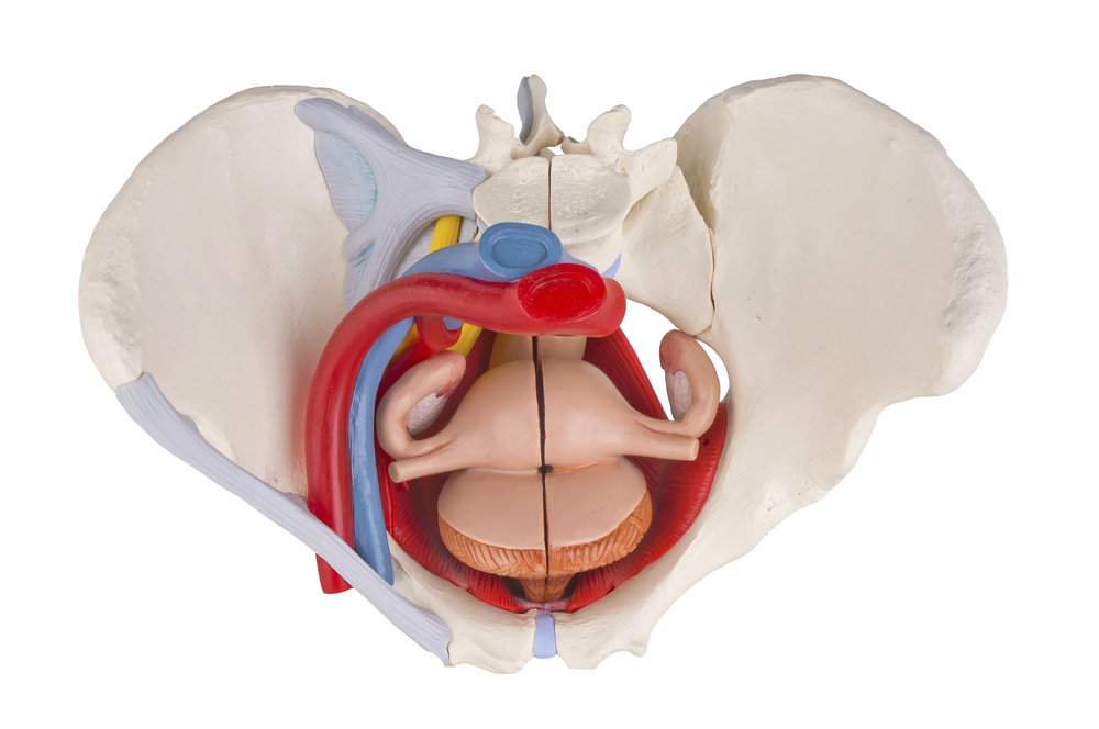
The pelvis is actually a bone ring found between the spine and lower limbs in the body. This protects the inner organs of the pelvis and the contents of the abdominal cavity. Leg muscles, back muscles, and abdominal muscles are attached to the pelvis.
Women's pelvic bones
The pelvic bone has a rotary joint attached to the femur and leg bones. This keeps the body upright, bending, and twisting and helps someone to walk or run.
Female pelvis are wider and lower than men, this is actually in accordance with women's needs during pregnancy and childbirth. The pelvic bone consists of three bones that are fused, namely the hip bone, the sacrum, and the coccyx.
Dialnsir of Health Line, the hip bone consists of:
- Ilium, which is the largest bone or main pelvic bone. This bone is on both sides of the spine and curves towards the front of the body. When holding your stomach, you will feel a prominent bone. It is the upper boundary of ilium called the iliac crest.
- Pubis, which is the bone in front of the hip bone close to the genitals. There is a combination of two pubic bones called the symphysis pubis, which is a very strong pubic joint. When giving birth, it becomes more flexible so that the baby's head can pass during labor.
- Ischium, which is the bone under the ilium and on the side of the pubis. This bone is thick because it is formed from two bones that are fused and circular. This is where the femur meets the pelvic bone and creates a hip joint.
Then there is the sacrum, which is a triangular bone located on the back of the pelvis consisting of five vertebrae that are fused. At the bottom of the sacrum there is a tailbone.

Pelvic canal
Rounded areas that are covered by pubic bones on the front and ischium on both sides behind it, are called the pelvic canal. This channel has a curved shape due to the difference in the size of the front and back created by the pelvic bone. This is a channel that babies must pass through when they are born.
In the pelvic area of women there are several important organs, such as:
- Endometrium (uterine lining), which is where the fertilized egg attaches
- The uterus, which is a hollow organ that is between the bladder and rectum (anus)
- Ovary (ovary), namely two female reproductive organs in the pelvis
- Fallopian tubes, which are channels that connect the ovary to the uterus.
- Cervix (cervix), which is the lower part of the uterus that forms an open channel into the vagina
Size and shape of the pelvis
After knowing the pelvic anatomy of a woman, you will understand that the size and shape of the pelvis greatly influences labor. Women with a broad pelvic size will find it easier to carry out normal labor.
Of course, the development of the pelvic bone is affected by food intake since childhood. Lack of important minerals such as iodine also makes the development of the pelvic bones become abnormal. In addition, women who experience stunting when they are small also tend to have narrower size.


