Contents:
- Medical Video: CT Scan
- Head CT scan procedure for diagnosing ischemic stroke
- Head CT scan procedure to diagnose hemorrhagic stroke
Medical Video: CT Scan
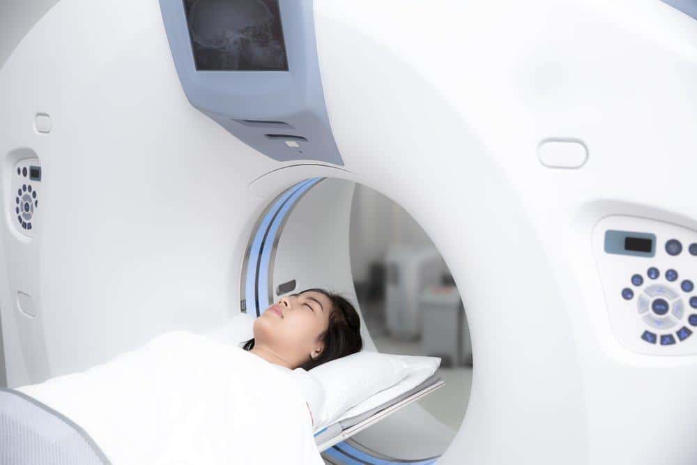
Computed tomography scan or a CT scan is a medical examination to read the condition of a person's body using X-ray technology and a computer at the same time. The results of this scan reading are clearer and more detailed than regular X-ray examinations. A head CT scan can be used by a doctor to find out which type of stroke you are experiencing - hemorrhagic stroke or ischemic stroke. Stroke is still the highest cause of death in the world. Therefore, scanning a head CT scan as early as possible can greatly help the doctor plan the treatment and recovery process.
Head CT scan procedure for diagnosing ischemic stroke
Ischemic stroke or blockage stroke is a type of stroke that occurs due to blockage of blood vessels in the brain. Ischemic stroke is responsible for 87 percent of total stroke cases.
Clinically, patients who experience a blockage stroke usually come with a complaint suddenly talking to pelo / hoarse / slurred / nasal sound and experience weakness on one side of the body. Ischemic stroke itself can be divided into three time phases.
- Acute phase (<7 days)
The first few hours after an ischemic stroke usually have not seen any abnormalities on the results of a CT scan of the head. Even if there is an abnormality, you may see parts of the brain that are darker than the same area on the opposite side. The dark part due to lack of oxygen intake, commonly called hypodense lesions, will be observed by the location and extent of the doctor.
- Subacute phase (8-21 days)
Over time after a stroke and not getting treatment as soon as possible, the color of the hypodens will darken more than the surrounding brain tissue.
- Chronic phase (> 21 days)
The CT scan of the head in this phase will show dark areas that are firmly demarcated with the surrounding brain tissue. This picture indicates that the area is a dead brain tissue. As a result, body functions regulated by the area will be disrupted and permanent if they do not get treatment from the start.
Head CT scan procedure to diagnose hemorrhagic stroke
Hemorrhagic stroke occurs when blood vessels in the brain leak or break. Hemorrhagic stroke accounts for about 13 percent of total stroke cases. This type of stroke starts from a weakened blood vessel, then ruptures and spills blood around it. Leaking blood builds up and blocks the surrounding brain tissue. Death or long coma will occur if bleeding continues.
Unlike an ischemic stroke, someone who has a hemorrhagic stroke usually comes with a more severe condition, among others loss of consciousness and vomiting to spray. On a CT scan the head of a hemorrhagic stroke patient can find the following:
- The area is white (hypersensitive lesion)
One of the initial points evaluated on the CT scan of the head of a hemorrhagic stroke patient is the presence or absence of a striking white area called a hyperdensic lesion. These lesions can ensure the doctor's diagnosis of bleeding in the brain. After that, the doctor will identify the location and the estimated amount of leaking blood in order to predict the next treatment.
- The right and left parts of the brain are not symmetrical
In addition to observing the presence of hiperdens lesions, the results of a scan of the CT scan of the head for hemorrhagic stroke will also be seen as a "space push effect" caused by the blood clots. The volume in the skull bones is limited, so that if there are additional blood clots there will be pressure on the surrounding area. The presence of pressure in the brain due to excess fluid will display the shape of both sides of the right and left brain that are not symmetrical in the reading.


