Contents:
Medical Video: British Heart Foundation - Your guide to MPS (Myocardial Perfusion Scan), heart disease test
Definition
What is a heart perfusion scan?
Cardiac perfusion scan is used to measure the amount of blood in the heart muscle when resting and during exercise. This scan is often done to find out the cause of pain in the chest. Can be done after a heart attack to see a part of the heart that does not get enough blood or find out the level of damage to the heart muscle due to a heart attack.
During the scan, the camera will take a picture of the heart after giving a special drug for this test (radioactive tracer) is given by infusion. Tracer moves through the blood and into the heart muscle. When tracer moves through the heart muscle, the area where the blood flow is good enough will absorb tracer. Areas that cannot absorb tracer may not get enough blood or damage from a heart attack. Two sets of images will be taken during a heart perfusion scan. A set is taken when you rest. Others are taken after your heart is working hard, either after exercising or after administering medication. The two images will then be compared.
This test is also known by other names, including myocardial perfusion scans, myocardial perfusion imaging, thalium scans, sestamibi heart scans, and nuclear stress tests.
When should I undergo a heart perfusion scan?
can heart perfusion be used to find out the cause of chest pain, or chest pain that occurs when exercising. This test can also be done to:
- shows the pattern of blood flow to the heart wall
- see if the heart (coronary) arteries are blocked and how severe the condition is
- determine the condition of an injury to the heart caused by a heart attack (myocardial infraction)
Prevention & warning
What should I know before undergoing a heart perfusion scan?
There are several reasons why you cannot take this test or your test results will not help your condition, including:
- severe heart attacks have occurred recently
- inflammation of the heart, such as myocardium or sarcoidosis
- injury to the heart muscle (cardiac contusion)
- weakened heart muscle
- tensed heart muscle (myocardial fibrosis)
- severe narrowing of the heart valve
- conditions that make it difficult for you to move, such as lung disease, arthritis, or problems with nerve muscles
- drugs, such as dipyridamole (Persantine) and pentoxifylline (Trental)
- electrolyte imbalance (specifically calcium, potassium, sodium and magnesium)
- pregnant or breastfeeding (except in an emergency)
Test results will be difficult to explain when a scan is performed on women with large breasts.
Drug stress tests will be carried out instead of sports stress tests for advanced adults and people with difficult exercise conditions, such as obesity or having chronic obstructive pulmonary disease, peripheral arterial disease, spinal cord injury, arthritis, or multiple sclerosis.
Process
What should I do before undergoing a cardiac perfusion scan?
In general there is not much preparation needed before this test. However, you will be asked not to eat or drink anything that contains caffeine before the test. In some cases, your doctor can advise you not to use the drug for several days before the scan. You may also be asked for a list of medicines you use on the day of the test. The local hospital will notify you if this rule applies to you.
How does the heart perfusion scan process?
Cardiac perfusion scans are usually performed in hospitals in the radiology department or doctor's office or outpatient clinic. This test is carried out by doctors and trained technicians in the field of nuclear medicine.
Resting scan
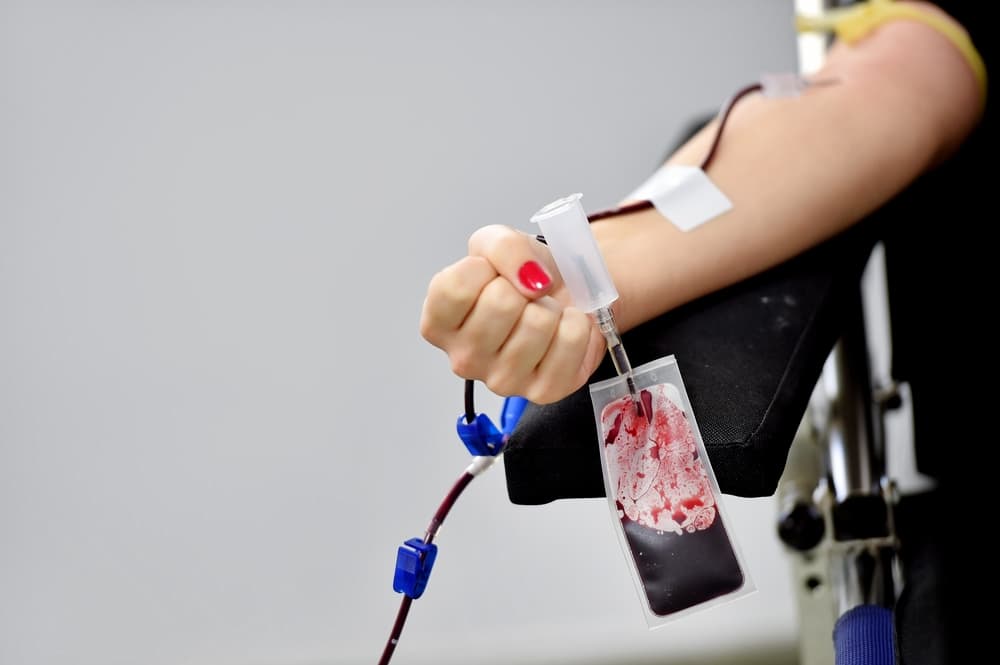
For resting scans you don't need to move, you will be asked to take off your clothes from the waist up, and you will be given hospital clothes to wear. The electrode will be attached to the chest to monitor your heart rate. Will be infused in the arm or hand. A small amount of radioactive tracer will be placed in IV.
You will lie on your back on a table with a large camera positioned above your chest. The camera records tracer signals when moving through your blood. The camera does not produce any radiation, so you are not exposed to additional radiation during the scan.
You will be asked to keep lying down every time the scan takes place, which takes between 5 to 10 minutes. The camera will move to take many pictures from different sides. Some scans will be needed.
The whole test takes 30 to 40 minutes, after which you can return to normal activities.
Stress scan using drugs
Stress scan is done in two parts. In many hospitals, the first picture is taken while resting. Then the second picture was taken after being given a drug such as adenosine, which gave a response to the heart as if it were after exercising. Sometimes stress scans are done first then resting scans are done the next day. Stress tests with drugs are usually used when a person cannot exercise for certain reasons.
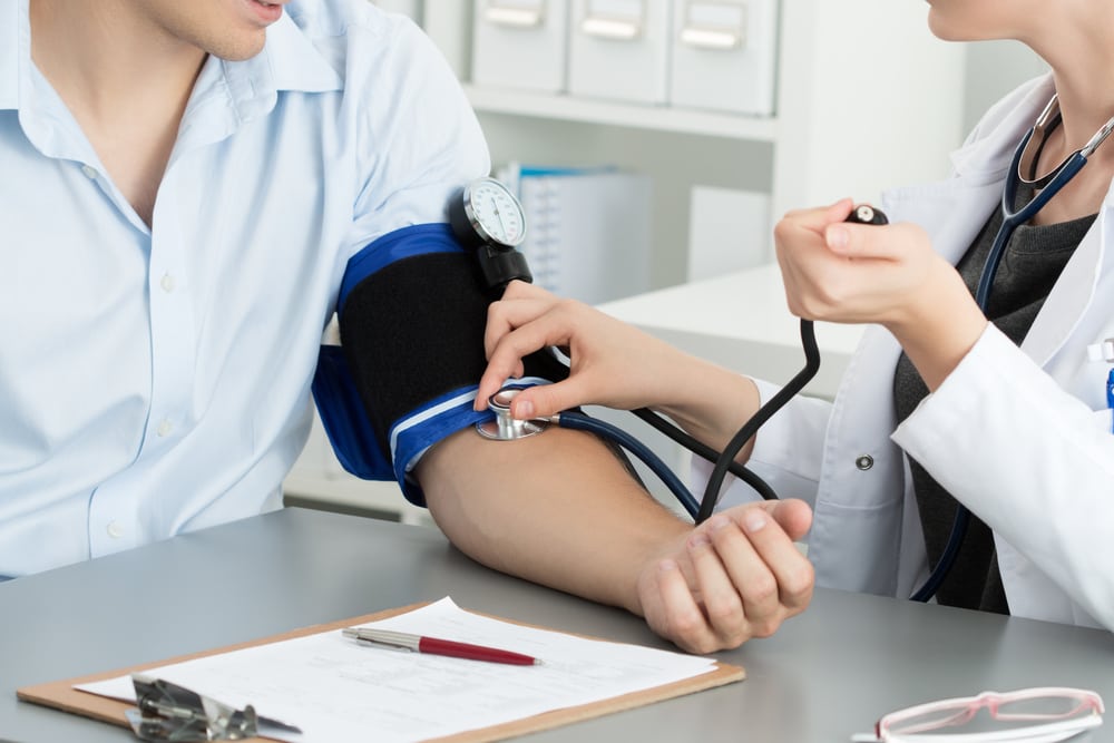
For this test, you will be asked to sit or lie on the examination table and you will be given an electrocardiogram (ECG or ECG), which takes about 5 to 10 minutes.
Then you will be given the drug via IV. You will feel headaches and dizziness and nausea due to the medication, but these side effects do not last long. An ECG and blood pressure measurements will be taken in addition. After the drug has reacted (about 4 minutes), a small amount of radioactive tracer (tracer) is given by infusion.
You will wait around 30 to 60 minutes. You will be asked to eat and drink something. Then you will lie on the table to do several scans. The camera records the tracer signal while moving through your blood. The camera does not produce any radiation, so you are not exposed to radiation during the scan.
Sometimes lots of pictures will be taken after you rest for 2 to 4 hours. Most people will return to their normal diet and activities after the last scan has been completed.
Stress scan with exercise
When stress scans with exercise, your heart rate will be checked with an electrocardiogram (ECG or ECG). Because ECG electrodes need to be attached to the chest to examine the heart, men will usually be bare chest and women usually only wear bras, or loose shirts.
Stress scan exercise is usually done in two parts. The first part, some pictures of resting will be taken then some stress images will be taken immediately after finishing the exercise. Sometimes a stress scan is done first then resting a scan the next day. In many hospitals, the first resting image is taken using one tracer type. The next image is taken using a different tracer after your heart is stressed because of exercise.
In this stress test, you will exercise on a treadmill or a static bicycle. Your heart rate will be checked during the test with standard electrocardiography. Your blood pressure will be checked using a blood pressure device placed on your arm. To learn more, see the topic of Exercise Electrocardiogram.
You will walk or pedal slowly and casually. Every minute, the speed will be increased. You will exercise until you need to stop or when you have reached the heart rate that feels right. At that time, you will be given a different tracer through an IV. You may continue to exercise in addition to 1 to 2 minutes so that the radioactive tracer circulation runs.
You will lie on a scan table. Each scan takes 5 to 10 minutes. The camera does not produce radiation, so you are not exposed to radiation during the scan. Sometimes more images will be taken after you rest for 30 minutes to 4 hours. In some hospitals, you are given a radioactive tracer a few hours after exercising and before the last picture.
What should I do after undergoing a cardiac perfusion scan?
You should wake up slowly to avoid dizziness or dizziness after lying on your back during the procedure. You will be instructed to drink enough fluids and urinate more frequently for 24 to 48 hours after the test to remove the remaining radionuclides from the body. Most people will return to their normal diet and activities after the scan is complete.
If you have questions relating to the process of this test, consult your doctor for a better understanding.
Explanation of Test Results
What do the test results mean?
Test results are usually available within 1 to 3 days. Scan for cardiac perfusion measures the amount of blood in the heart muscle at rest and during exercise. The result is:
- normal, if the radioactive tracer evenly flows in your heart muscle
- abnormal, if there is an abnormal absorption section of the tracer. This indicates that some parts of the heart muscle do not get enough blood (ischemia). This can mean there is damage to the heart or coronary artery disease
Hello Health Group does not provide medical advice, diagnosis or treatment.