Contents:
- Medical Video: Nephron Function
- Anatomy of the human kidney
- 1. Cortex (Cortex)
- 2. Medulla (medulla)
- 3. Kidney pelvis (renal pelvis)
- Get to know nephrons, parts of the kidneys that filter blood
- 1. Malphigi's body
- 2. Kidney tubules
- Stages of urine formation
- The first stage
- Second stage
- Third phase
- Fourth stage
Medical Video: Nephron Function
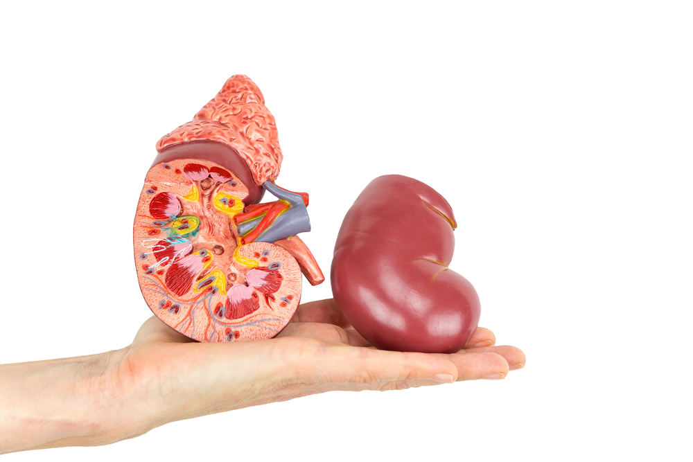
Kidney is one of the important organs of the body that functions to filter blood. Everyone has a pair of kidneys in his body. To find out more details, here are reviews about kidney anatomy.
Anatomy of the human kidney
The kidneys are located along the muscular wall of the back (posterior muscle) of the abdominal cavity. The shape of the kidneys resembles peanuts that are about the size of a hand. The kidneys are equipped with a pair of ureters, a bladder and urethra that carry urine out.
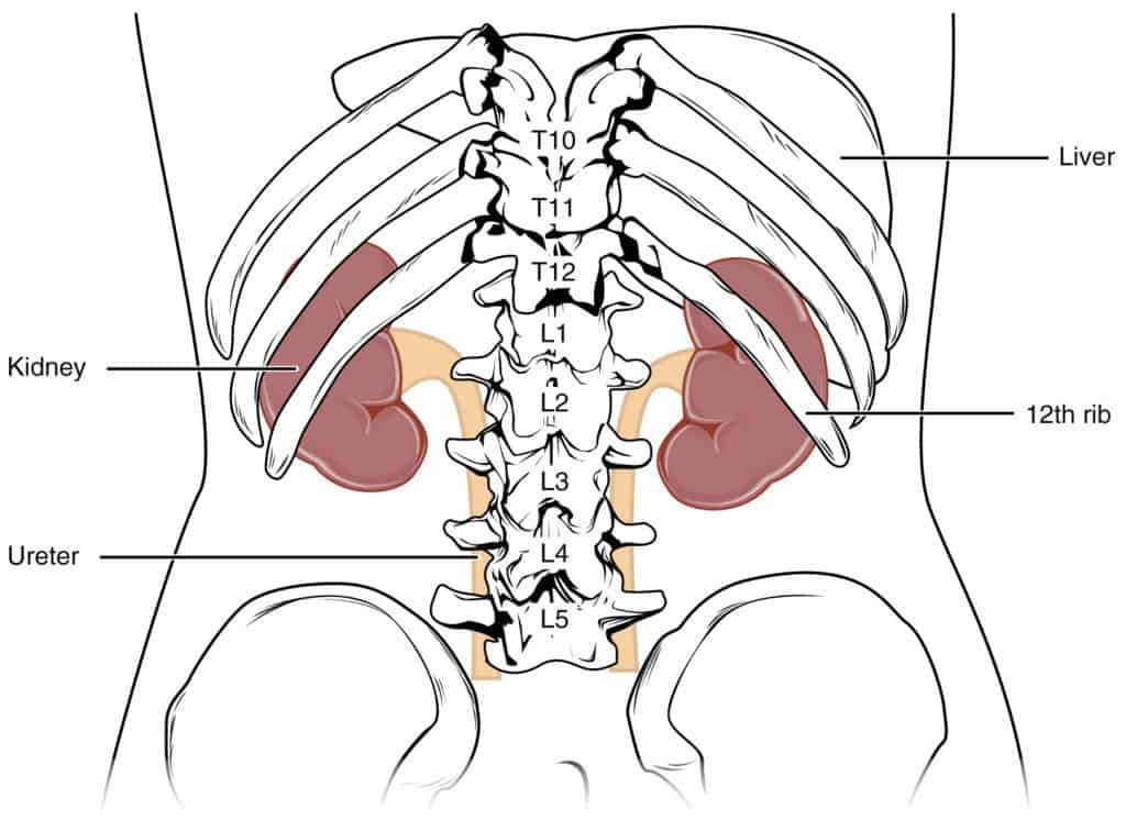
Humans have a pair of kidneys whose left part is located slightly higher than the right kidney, because of the presence of the liver that urges the right kidney. The kidneys are also protected by ribs and back muscles. In addition, adipose tissue (fat tissue) surrounds the kidneys and acts as a protective kidneys.
In general, the anatomy of the human kidney is divided into three parts from the outermost to the deepest, namely the renal cortex, renal medulla, and renal pelvis.
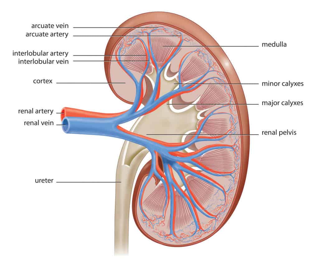
1. Cortex (Cortex)
The kidney cortex is the outermost part of the kidney. The outer edge of the kidney cortex is surrounded by kidney capsules and fat tissue, to protect the inside of the kidney.
2. Medulla (medulla)
The kidney medulla is a smooth and deep kidney tissue. The medulla contains the arches of Henle and the kidney pyramid, which are small structures with nephrons and tubules.
This tubule transports fluid to the kidney which then moves away from the nephron to the part that collects and transports urine out of the kidney.
3. Kidney pelvis (renal pelvis)
The kidney pelvis is a funnel-shaped space in the innermost part of the kidney. This serves as a pathway for fluid on the way to the bladder. The first part of the kidney pelvis contains calyces. This is a small cup-shaped chamber that collects fluid before moving into the bladder.
Hilum is a small hole located on the inside of the kidney, where it curves inward to create a different bean-like shape. Kidney pelvis passes, and:
- The kidney arteries carry oxygen-rich blood from the heart to the kidneys for the filtration process.
- Kidney veins, bring filtered blood from the kidneys back to the heart.
A ureter is a muscle tube that pushes urine into the bladder.
Get to know nephrons, parts of the kidneys that filter blood
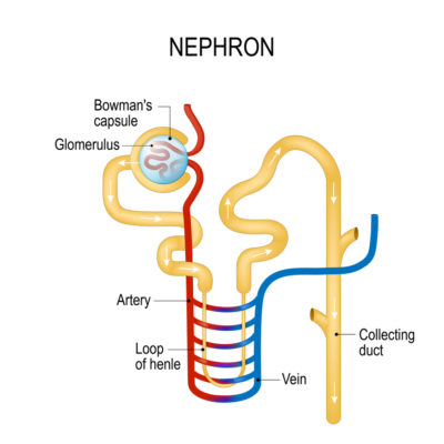
Nephrons are part of the anatomy of the kidneys that are responsible for blood filtration. Nephrons take blood, metabolize nutrients, and help circulate filtered waste products.
Nephrons extend across the cortex and renal medulla area. Each kidney has about one million nephrons, each of which has its own internal structure. Here are parts of the nephron:
1. Malphigi's body
After blood enters the nephron, blood enters the malpighi body (kidney corpus). Malphigi's body contains two additional structures, namely:
- Glomerulus, a group of capillaries that absorb protein from blood through the malphigi body.
- Bowman Capsule.
2. Kidney tubules
Kidney tubules are a series of tubes that start after the Bowman capsule and end in the collecting tubules (collecting duct). Each tubule has several parts:
- Proximal tubule is the tubule closest to the glomerulus, the shape of this tubule is convoluted. Function to absorb water, sodium, and glucose back into the blood.
- Henle (loop of henle) curvature is part of the kidney tubules which form a downward arch, and are located between the proximal and distal tubules. Function to absorb potassium, chloride and sodium into the blood.
- Distal Tubules is a tubule which is at the end of a series of renal tubules with a convoluted shape. Serves to absorb more sodium into the blood and take potassium and acid.
The waste or liquid filtered from nephrons is passed into the collecting tubules, which direct urine to the renal pelvis. Pelvic kidney with ureter allows urine to flow to the bladder for excretion.
Stages of urine formation
The kidneys are organs that are responsible for filtering blood and making urine. Every day, two kidneys filter around 120-150 liters of blood to produce about 1-2 liters of urine, consisting of waste and extra fluid. Urine flows from the kidney to the bladder through the ureter, which is on each side of the bladder, to be stored.
Here is how the kidneys work when filtering blood and producing urine:
The first stage
The process of urine formation begins with screening (filtration) of blood, which is carried out by the glomerulus in blood flowing from the aorta through the kidney arteries to the Malpighi's body.
This residual filtering product is called primary urine, which contains water, glucose, salt and urea. These substances will enter and be temporarily stored in Bowman's capsule.
Second stage
After the primary urine is stored temporarily in Bowman's capsule, then it will go to the collecting channel. On the way to this collecting channel, the process of urine formation through the stages of reabsorption.
Substances that can still be used such as glucose, amino acids, and certain salts will be absorbed again by the proximal tubules and arches of Henle. Reabsorption of the primary urine will produce secondary urine. Secondary urine is characterized by a high content of urea.
Third phase
The last urine formation process is the release of substances (augmentation). Secondary urine produced by the proximal tubule and Henle's arch will flow to the distal tubule.
Urine secretion will pass through blood capillaries to release substances that are no longer useful to the body. Next, the actual urine is formed.
Fourth stage
When the bladder meets capacity, the signal sent to the brain tells someone to go to the toilet immediately. When the bladder is empty, urine flows out of the body through the urethra, which is located at the bottom of the bladder.
In general, the kidneys are useful for maintaining homeostasis (balance of various bodily functions) in the body and help control blood pressure.
The kidneys maintain balance in electrolytes, acid bases, and fluids in the blood. The kidneys remove nitrogenous waste from the body (creatinine, urea, ammonia) and maintain important substances that the body needs to function properly.
In addition, the kidneys also produce the hormone erythropoietin which stimulates the production of red blood cells and enzymes.