Contents:
- Medical Video: Radiography Examination: What You Need To Know For The ARRT Radiography Exam Part 1
- Radiological examination is an important procedure for diagnosing disease
- Division of radiology
- 1. Diagnostic radiology
- 2. Radiology intervention
- When to go to a radiologist?
- Side effects of examination with imaging technology
- Technical preparation before radiology examination
Medical Video: Radiography Examination: What You Need To Know For The ARRT Radiography Exam Part 1
Radiology is a branch of medicine to know the inside of the human body using imaging technology, both electromagnetic waves and mechanical waves. Doctors who specialize in radiology are referred to as radiologists or radiologists.
Radiologists themselves act as expert consultants whose duty is to recommend the required examination, interpret medical images from the results of the examination, and use the results of tests to direct treatment that is appropriate to the patient's condition. One of the most well-known types of radiological examinations is X-rays using X-rays. Even so, radiological examination is not only that. Check out other important information about radiology in the medical world below.
Radiological examination is an important procedure for diagnosing disease
In the world of medical radiology plays a very important role. Without imaging technology, the disease will be difficult to diagnose and the existing treatment will not work optimally. As a result, more and more people are sick and die because the disease is not diagnosed early.
The key is simple, the earlier the disease is diagnosed, the greater the chance for patients to experience recovery.
Some conditions that can be known through radiological examination are:
- Cancer
- Tumor
- Heart disease
- Stroke
- Lung abnormalities
- Disorders of bones and joints
- Disturbances in blood vessels
- Disorders of liver and kidney function
- Disorders of the thyroid gland and lymph nodes
- Disorders of the digestive tract
- Disorders of the reproductive tract
Division of radiology
Radiology can be divided into two different fields, namely:
1. Diagnostic radiology
Radiology diagnosis helps doctors and health staff to see the structure in your body using imaging technology. This is done for:
- Know the condition of the inside of the patient's body
- Diagnose the causes of symptoms that patients complain about
- Monitor how well the patient's body responds to treatment or treatment
- Do screening for various diseases, such as cancer, heart disease, lung disease, stroke, joint and bone disorders, epilepsy, strokes, infections, disorders of the thyroid gland, and so on.
The most common types of diagnostic radiological examinations include:
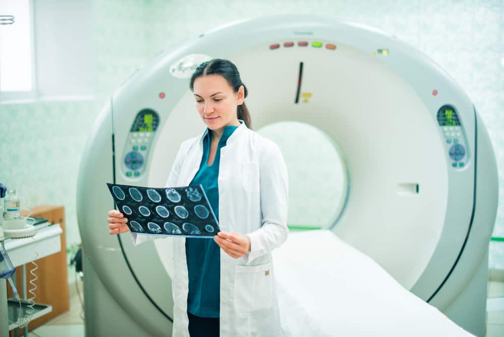
- Computed tomography, as known as computerized axial tomography (CT / CAT) scans, including CT angiography
- Fluoroscopy
- Magnetic resonance imaging (MRI) and magnetic resonance angiography (MRA)
- Mammography
- Nuclear examination, like bone scan, thyroid scan, and heart thallium stress tests
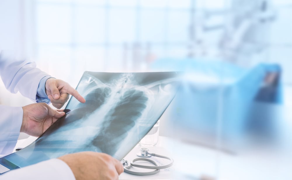
- X-rays
- Positron emission tomography, also called PET imaging, PET scan, or PET-CT when combined with CT
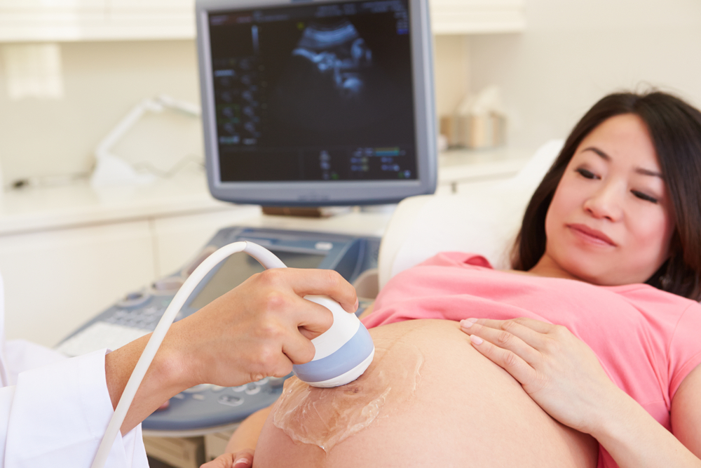
- Ultrasound (USG)
2. Radiology intervention

Interventional radiology allows doctors to perform minimally invasive (minimally invasive) medical procedures to diagnose and treat diseases. Guided from images obtained through imaging technology, doctors can insert catheters, cameras, cables, and other small instruments into certain parts of the patient's body. Compared to medical procedures that have to involve open surgery, minimally invasive techniques have a lower risk and faster recovery time.
Doctors who are experts in this field are often involved in the treatment of cancer, heart disease, blockages in the arteries and veins, uterine fibroids, back pain, liver and kidney function disorders, and so on.
Examples of interventional radiological procedures include:
- Angiography, angioplasty and blood vessel ring placement
- Embolization to stop bleeding
- Chemotherapy through arteries
- Biopsy of needles from different organs, such as the lungs and thyroid gland
- Breast biopsy, guided by technique stereotactic or ultrasound
- Placement of feeding tubes
- Catheter installation
When to go to a radiologist?
Before finally someone is recommended to consult a radiologist, there are several stages of examination that must be undertaken. In the initial stage, a patient will first undergo an examination at a general practitioner. If at this stage the general practitioner finds some symptoms that lead to certain diseases that require further examination, the general practitioner will refer the patient to the radiologist. The same thing can happen if you see a specialist.
Later, the radiologist will carry out further examinations to establish the initial diagnosis made by general practitioners or specialists. To ensure a diagnosis, the radiologist will usually carry out the most appropriate examination to diagnose your complaint.
The results of examinations carried out by a radiologist can provide additional information to a general practitioner or specialist who provides a referral to a radiologist.
Side effects of examination with imaging technology
Although examinations carried out with imaging technology are fairly safe, there are still some risks of possible side effects. Some of them are like:
- Patients can experience nausea, vomiting, dizziness, itching on the skin, feeling a metallic sensation in the mouth due to contrast fluid injected into the body. In rare cases, contrast fluids can also cause blood pressure to experience a drastic decline, anaphylatic shock, and a heart attack.
- X-rays can affect the development and growth of infants and fetuses.
- There is a study that states that CT scan procedures can increase the risk of cancer and can damage DNA, especially in pediatric patients. However, this risk is very small to occur, possibly only 1 in 2,000 cases. So, CT scans are still considered a safe enough examination and can help doctors evaluate the condition of their patients.
- Contrast fluids can cause allergies in some people.
Technical preparation before radiology examination
Basically, each procedure requires different preparation. Before undergoing a radiological examination, the doctor will usually tell what the patient must prepare. Here are some of the general things that doctors often recommend:
- Wear comfortable and loose clothing to make it easy to open when the inspection takes place. Even so, some hospitals will provide special clothes for patients to wear.
- Removing jewelry, watches, glasses, or metal-containing instruments on the body. If you have metallic implants in the body, such as the installation of a heart ring, or a nut in the bone, immediately report it to the doctor. The reason is, these objects will block X-rays to penetrate into the body.
- The patient may be asked by the doctor not to eat and drink for several hours before the examination is done.