Contents:
- Medical Video: Anatomy of the Heart
- What is the anatomy of the human heart like?
- Bile duct
- Blood vessel
- Lobules
Medical Video: Anatomy of the Heart
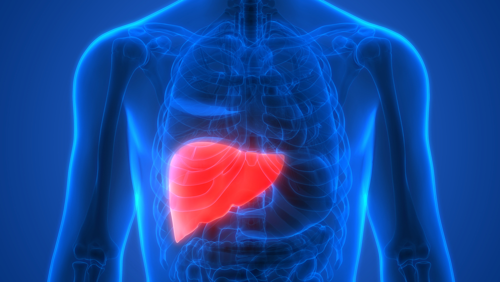
The liver is a vital organ that plays an important role in the digestive and metabolic systems, storing the body's nutrients, as well as the body's immunity. But there are still many who do not know what and the functions of each part of the heart. Come on, see the following anatomical description of the liver.
What is the anatomy of the human heart like?
Maybe all this time you consider if the liver has a shape like ‘love ’ or 'ivy leaves'. Even though an organ weighing no more than 1.5 kg is shaped like a triangle that is located on the right side of the abdominal cavity and below the diaphragm.
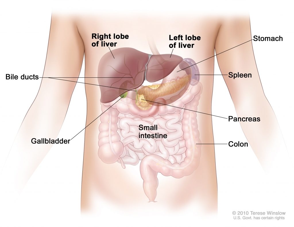
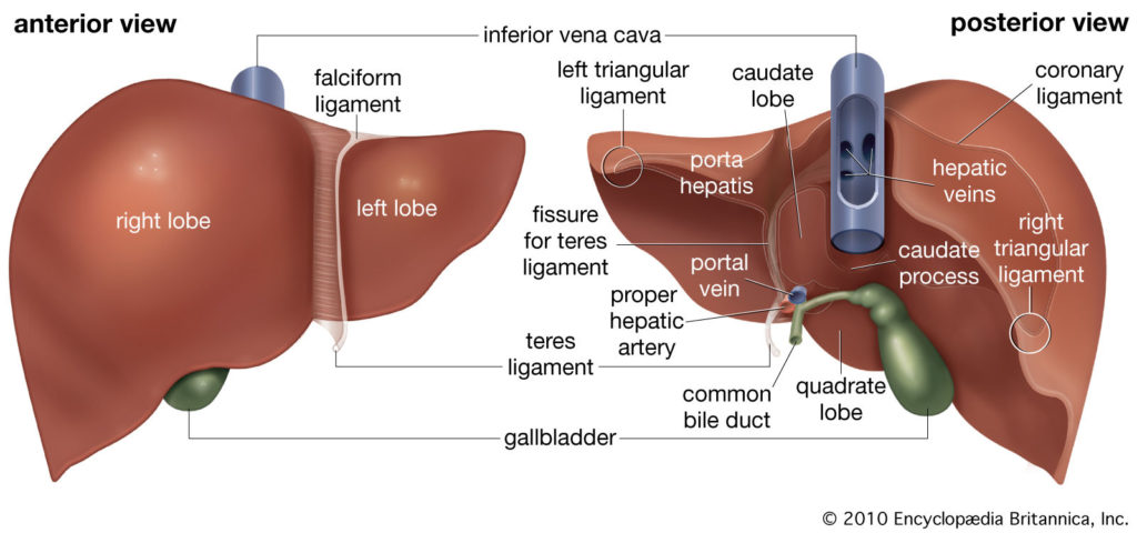
The anatomy of the human heart when viewed with the naked eye, consists of four lobes (parts) of different sizes.
- The right lobe is the largest part of the liver that is 5 to 6 times larger than the left lobe.
- The left lobe is the part of the heart that has a more pointed and smaller shape than the right lobe. The left and right lobes are separated by the falciform ligaments.
- The caudate lobe is smaller than the two previous lobes, located extending from the back of the right lobe and wrapping the main reverse vein (inferior vena cava).
- The quadratic lobe is lower than the caudate lobe and is located from the back of the right lobe to the gallbladder. The squared lobe and caudate are rarely seen in anatomical images because they are located behind the left and right lobes.
Bile duct
The bile duct is a channel that connects the liver and gallbladder (bile storage). Bile is a substance produced by the body to help digest fat and will be stored in the gallbladder. Furthermore, the bile duct meets the larger left and right hepatic ducts, which carry bile from the left and right lobes of the liver.
The two hepatic channels then merge to form a channel to drain all bile from the liver. Most of the bile produced by the liver is transferred to the storage bag, until it is used for the digestive process.
Blood vessel
The blood supply from the liver is unique compared to other organs because there is a hepatic portal vein system. Blood from organs such as the spleen, pancreas, gallbladder and intestine collects in the hepatic portal vein. Then from here, blood is sent to the liver which will be processed before it is ready to continue.
Blood from the liver will gather in the hepatic vein and lead to the vena cava and then return to the heart. Just like other organs, the human heart also has its own system of arteries and arterioles which produce blood containing oxygen for their tissue needs.
Lobules
The internal structure of the heart is composed of about 100,000 hexagonal liver cells, known as lobules. Each lobule consists of a central blood vessel surrounded by six hepatic veins and six hepatic arteries. These blood vessels are connected by many tortuous small blood vessel channels called sinusoids.
Each sinusoid has two main types of cells, namely cells and hepatocyte cells. Kupffer cells are cells derived from white blood cell tissue and function to destroy foreign substances or dead cells. In the liver, kuppfer cells are responsible for capturing and breaking old red blood cells and passing them to hepatocyte cells.
While hepatocyte cells are the cells that line the sinusoid and form most of the cells in the liver. Hepatocytes play an important role because they do most of the liver's functions, namely digestion, metabolism, bile storage and production.