Contents:
- Medical Video: Anatomy of the Eye
- Anatomy of the human eye and its function
- 1. Cornea
- 2. The front chamber (anterior chamber)
- 3. Sklera
- 4. Iris and pupil
- 5. Lens
- 6. Choroid and conjunctiva (conjunctiva)
- 7. Vitreous
- 8. Retina and optics
Medical Video: Anatomy of the Eye
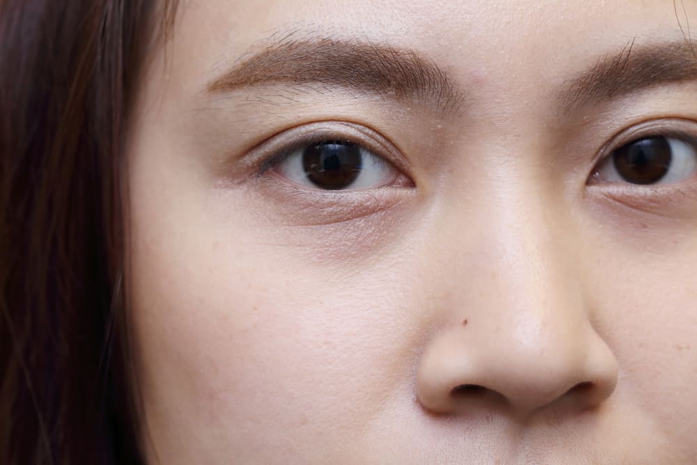
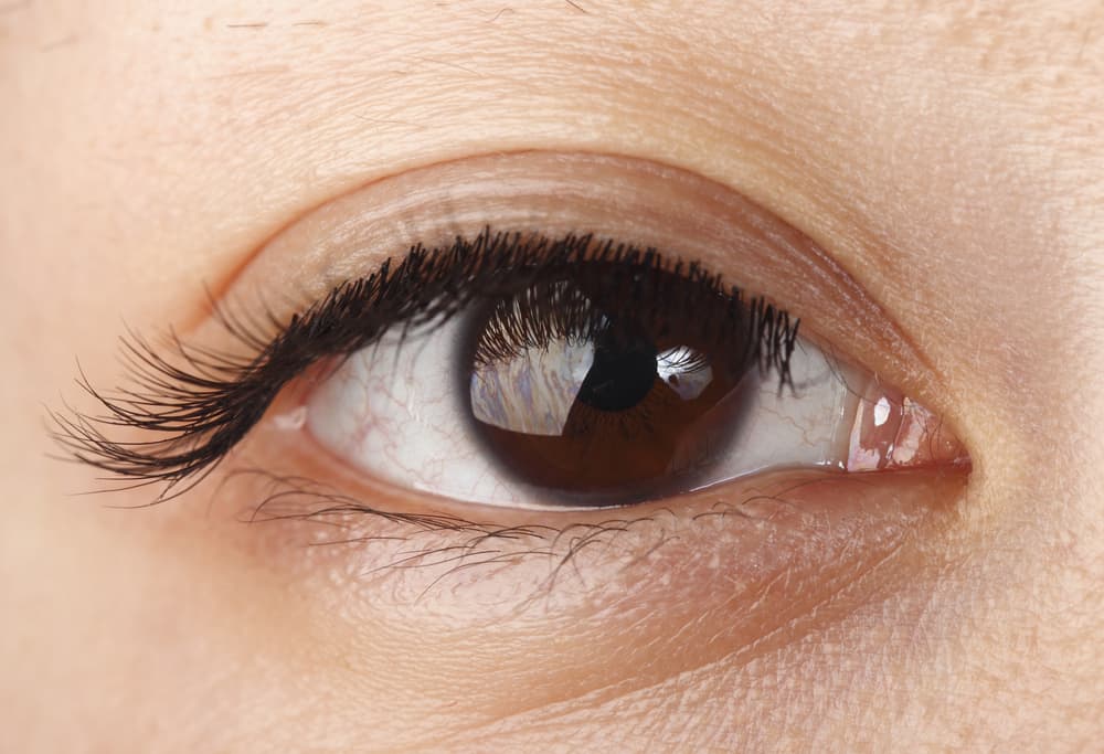
The eye is one of the most important organs of the body. However, there are still many who don't know about the important parts of the anatomy of the eye. To get a brief explanation of the anatomy of the eye and the function of each part, see the reviews below.
Anatomy of the human eye and its function
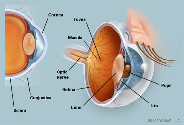
1. Cornea
The cornea is a transparent dome-shaped tissue that forms the very front of your eyes. The cornea serves as a window and as a way of entering light into your eyes. Thanks to the cornea, your eyes also begin the process of regulating the process of light so that you can see the words and images clearly.
The cornea serves to provide 65-75 percent of the focus of your eye's strength. You also need to be careful to maintain the health of your cornea. Because inside the cornea contains many nerve endings that make it very sensitive and an important part in the pathway of the entry of light into the eye.
2. The front chamber (anterior chamber)
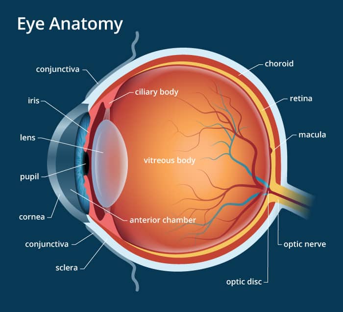
The front chamber is a jelly-like bag behind the eye cornea, in front of the lens. The sac also known as the termanterior chamber this (see in the picture) contains aqueous humor that helps bring nutrients to the eye tissue. This aqueous humor serves as a counterweight to the pressure inside the eye.
Your eye health depends on the production process and fluid flow in the front chamber. If there is a problem, this can cause pressure problems in his eyes, such as glaucoma.
3. Sklera
Sklera is a hard white membrane with fibrous tissue that covers your entire eyeball (all the way around), except the cornea. Inside is a muscle attached to move the eye attached to the sclera.
4. Iris and pupil
The iris and pupil are parts of the eye anatomy that are interconnected with one another. Iris is a ring-shaped membrane inside the eye that surrounds the hole in the middle. Now the hole in the middle is what is called the pupil. Pupils are muscles that can be closed and open or shrink and enlarge.
Iris functions to regulate the amount of light that enters the eye and adjusts to the pupil opening. When exposed to bright light, the iris will close (or narrow) and make the pupils open smaller to limit the amount of light that enters your eyes.
In addition, the iris determines the color of your eyes. People with brown eyes have high pigmented irises, while people with blue or light eyes have little pigments.
5. Lens
The lens is a transparent and flexible tissue that is located just behind the iris and pupil. This is one of the second parts of your eye, after the cornea. The function of the lens is to help focus light and images on your retina.
Because the lens of this eye is flexible and elastic, its formation can turn into a curve and focus on objects around, people who are nearby or from a distance. This lens gives 25-35 percent of your eye's focus strength.
As we age, one of the important parts of the eye's anatomy can lose its elasticity and the ability to capture objects in focus. This is commonly referred to as presbyopia or old eyes, which is a visual impairment that many older people experience.
6. Choroid and conjunctiva (conjunctiva)
Choroid is a dark brown membrane that has many blood vessels in it. Its position is located between the sclera and retina. Choroid serves to supply blood and nutrients to the retina and to all other structures in the anatomy of the eye. While the conjunctiva is a thin layer of tissue that covers the entire front of your eye, except for the cornea.
7. Vitreous
Unlike the aqueous humor that is present in front of the lens of the eye, the vitreous humor is located behind the eyepiece. Vitreous is a jelly-like substance that fills the inside of the back of the eye. Over time, the vitreous becomes thinner and can be released from the back of the eye.
If your eyesight looks like there is a white cloud floating or the light blinks, immediately see an ophthalmologist. Because the separate vitreous substances can cause a hole (a condition called the macular hole) to develop in the retina.
8. Retina and optics
Retina is a network that is sensitive to light. This retina covers the inner surface of the eye. Cells in the retina can turn incoming light into electrical impulses. This electrical impulse is carried by the optic nerve (which resembles your television cable) to the brain, which finally interprets it as the image or object you see.
Whereas the macula is a small sensitive area in the middle of the retina which provides clear central vision. The Fovea is located at the center of the macula and its function is to provide the sharpest vision in your eyes.