Contents:
- Medical Video: Anatomy of the Heart
- Parts of the heart and their functions
- Pericardium
- Porch
- Booth
- Valve
- Blood vessel
- Heart cycle
- Various diseases that can cause heart problems
Medical Video: Anatomy of the Heart
The heart is a vital part of your body that is responsible for receiving and pumping blood throughout the body. This organ has a size that is slightly larger than your fist, which is around 200-425 grams. The location of the heart is between the lungs, in the middle of the chest, precisely on the left back of the breastbone. For more details, the following is the anatomy of the heart as well as an explanation of its parts and functions.
Parts of the heart and their functions
Intrigued how can an organ which is no more than a fist be able to trash blood throughout the body? Following is a brief description of heart anatomy and its function.
Pericardium
The heart is in a fluid-filled cavity called the pericardial cavity. This wall and layer of the pericardial cavity is called the pericardium. Pericardium is a type of serous membrane that produces serous fluid to lubricate the heart during pulsing and prevents painful friction between the heart and surrounding organs. This part also serves to support and hold the heart in place. The heart wall consists of three layers, namely epicardium (outermost layer), myocardium (middle layer), and endocardium (inner layer).
Porch
Porch or also called the atrium is the upper part of the heart which consists of the right and left porch. Right porch serves to receive dirty blood from the body carried by blood vessels. While left porch serves to receive clean blood from the lungs. Serambi has thinner and not muscular walls because its job is only as a recipient of blood.
Booth
Just like the porch, cubicle or also called the ventricle is the lower heart part which consists of the right and left parts. Right booth serves to pump dirty blood from the heart to the lungs. Meanwhile, left booth serves to pump clean blood from the heart throughout the body. The wall of the cubicle is much thicker and more muscular than the porch because it works harder to pump blood from the heart to the lungs and throughout the body.
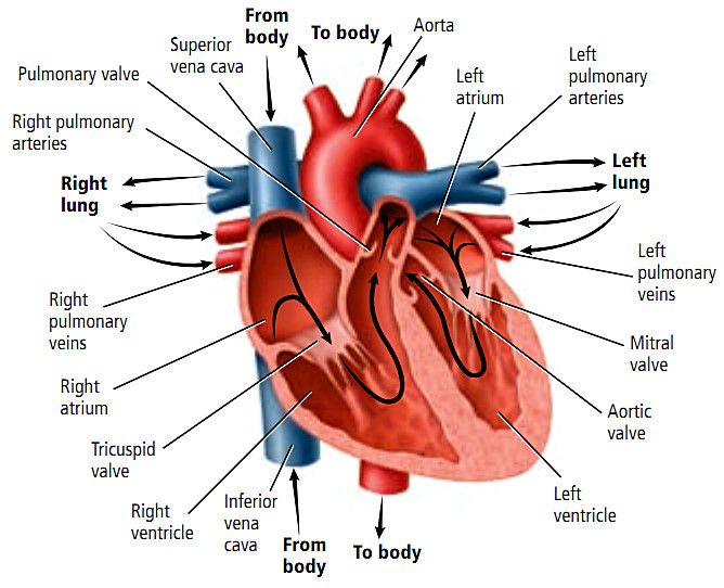
Valve
The heart has four valves that keep blood flowing in one direction, namely:
- Tricuspid valve, regulate blood flow between the right foyer and the right chamber.
- Pulmonary valve, regulates blood flow from the right chamber to the pulmonary artery which carries blood to the lungs to extract oxygen.
- Mitral valve, flowing oxygen-rich blood from the lungs flows from the left porch to the left ventricle.
- Aortic valve, paving the way for oxygen-rich blood to be passed from the left ventricle to the aorta (the largest artery in the body).
Blood vessel
There are three main blood vessels in the heart, namely:
- Arteries, carrying oxygen-rich blood from the heart to other body parts. Arteries have walls that are elastic enough to keep blood pressure consistent.
- Veins, this one blood vessel carries oxygen-poor blood from the entire body to return to the heart. Compared to arteries, veins have thinner vessel walls.
- CapillaryThis blood vessel is responsible for connecting the smallest arteries with the smallest veins. The walls are so thin that it allows blood vessels to exchange compounds with surrounding tissues, such as carbon dioxide, water, oxygen, waste, and nutrients.
Heart cycle
Heart cycle is a sequence of events that occur when the heart beats. The following are two phases of the heart cycle, namely:
- Systole, heart muscle tissue contracts to pump blood out of the ventricles.
- Diastolthe heart muscle relaxes when filling the blood in the heart
Blood pressure increases in the main artery during ventricular systole and decreases during ventricular diastole. This causes 2 numbers associated with systolic blood pressure is a higher number and diastolic blood pressure is a lower number. For example, a blood pressure of 120/80 mmHg describes systolic pressure (120) and diastolic pressure (80).
Various diseases that can cause heart problems
Heart disease is a term that includes any heart problems. The following are various disorders of the heart, including:
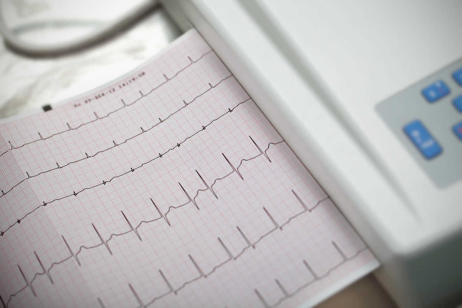
- Arrhythmia, irregular heartbeat.
- Coronary artery, occurs due to a buildup of plaque that clogs blood flow in the coronary arteries.
- Heart failure, occurs when the heart is unable to pump blood throughout the body effectively.
- Dilated cardiomyopathy, occurs when the heart chambers widen due to weakening of the heart muscle so that it cannot pump blood properly.
- Hypertrophic cardiomyopathy, is a genetic disorder that occurs when the left ventricle thickens making it difficult for blood to be pumped out of the heart.
- Pulmonary stenosis, occurs when the pulmonary valve is difficult to open, making it difficult for the heart to pump blood from the right chamber to the pulmonary artery.